Research Overview
Click on different image panels for the overview
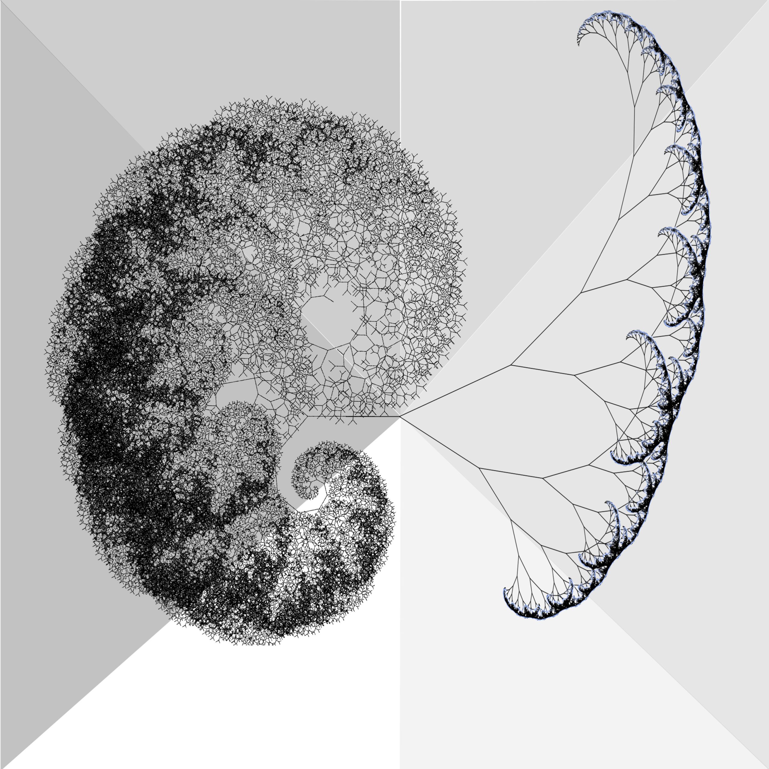
Advanced Imaging
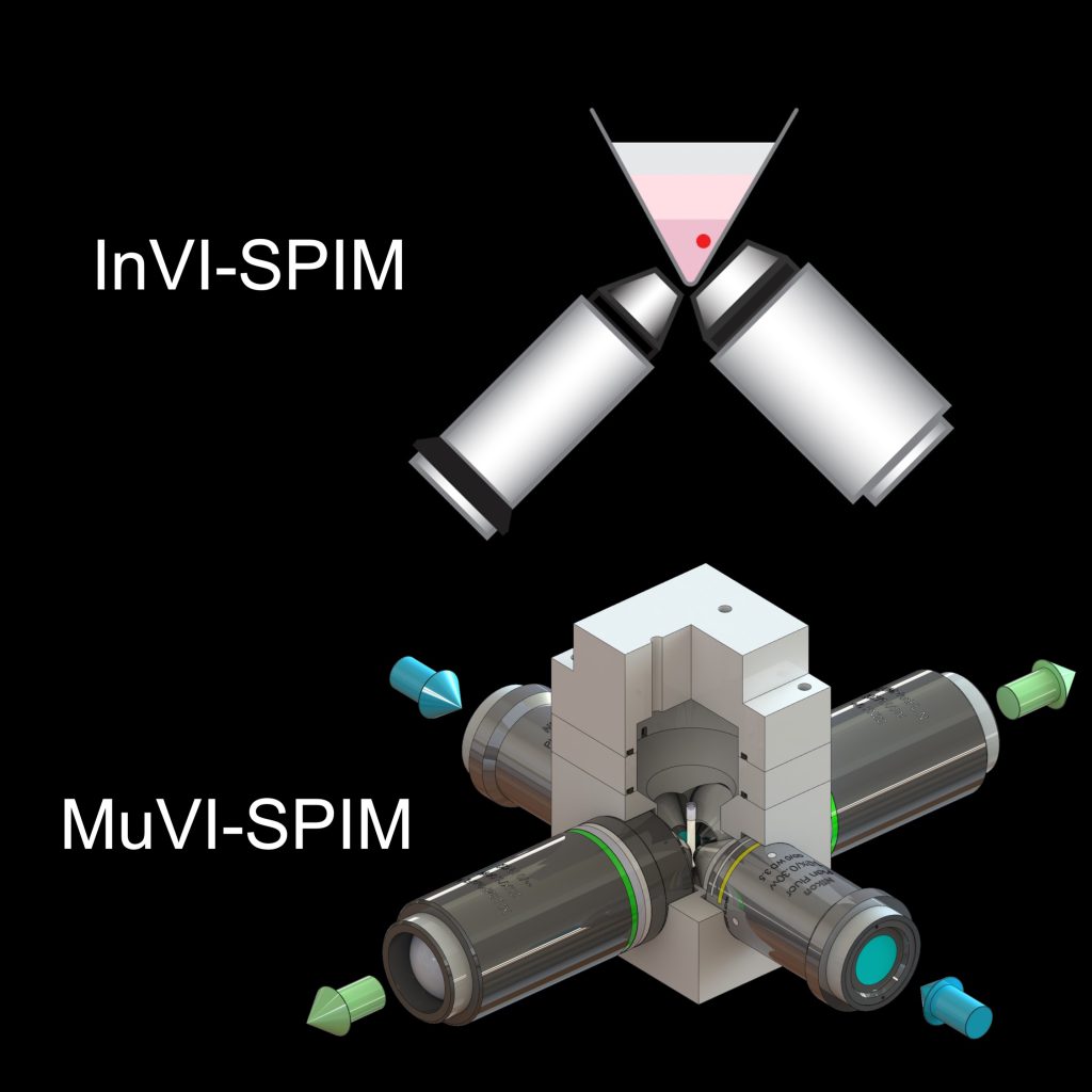
Engineering solutions for long-term live imaging of complex 3D samples such as developing embryos using light-sheet microscopy.
Bondarenko V, et al., EMBO J. (2023)
Bondarenko V, et al., EP23162464.4 (2023)
Bioengineering
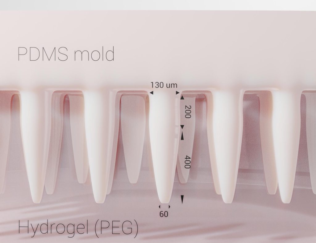
Ex vivo Engineering Uterine micro-Environment with topographically patterned hydrogels (3E-uterus) for development of the whole mouse embryo during implantation
Bondarenko V, et al., EMBO J. (2023)
Stem Cell Embryo Models
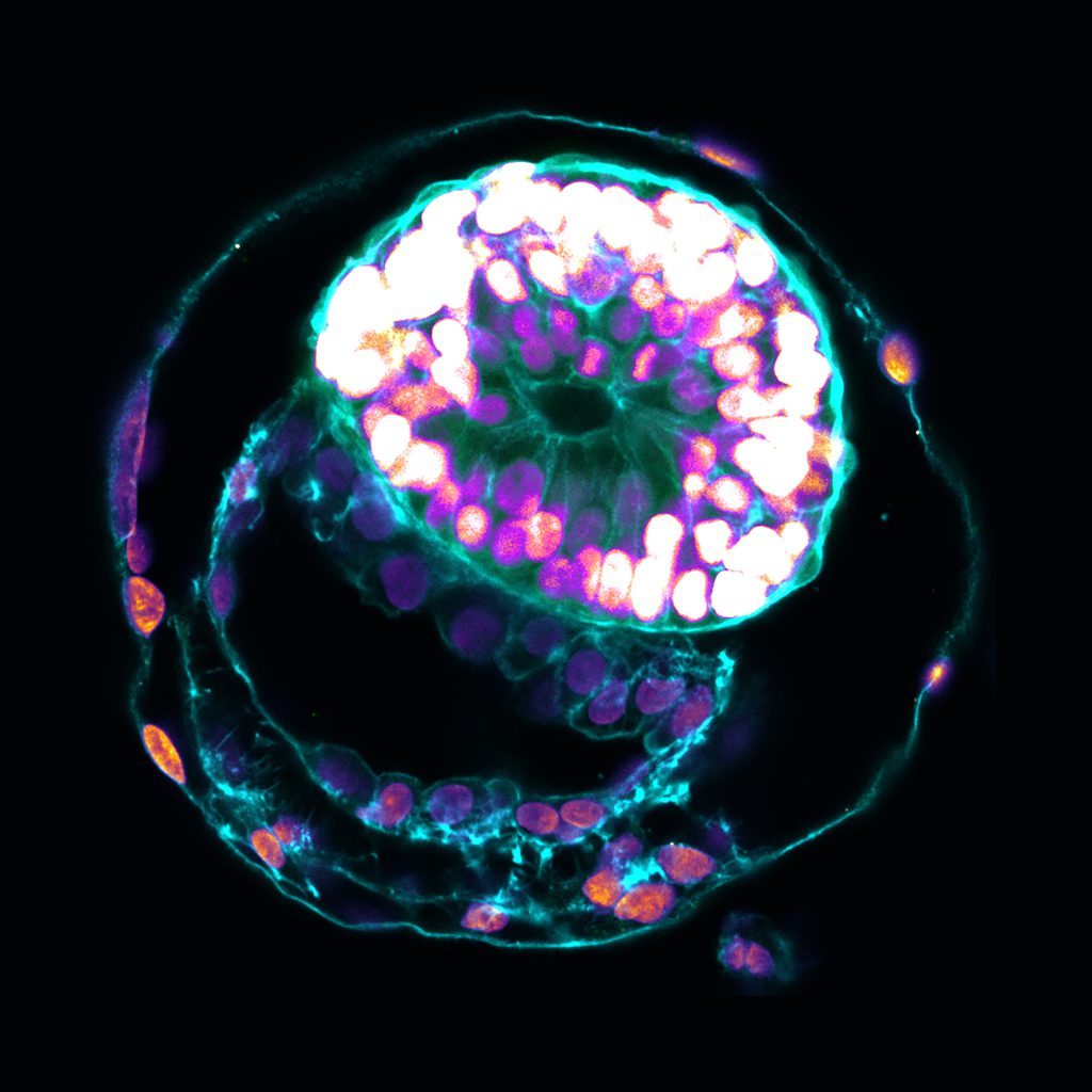
Using human embryonic stem cells to model early post-implantation human embryo development.
Oldak B*, Wildschutz E*, Bondarenko V*, et al., Nature (2023)
In Vivo Development
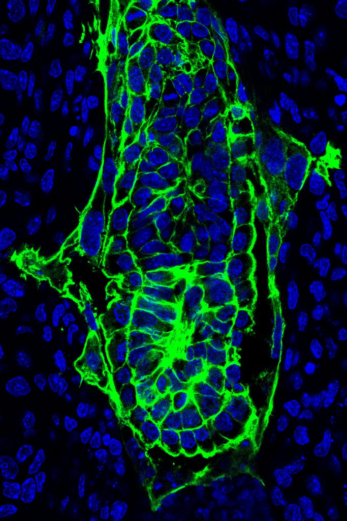
Dissecting in vivo uterine tissue context and development using various imaging and spatial multiomics techniques.
Theoretical modeling
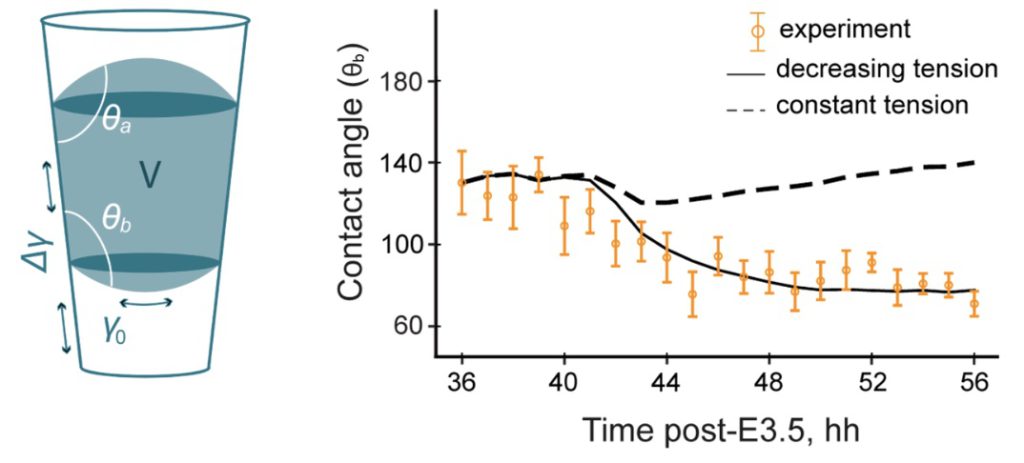
Applying concepts from theoretical physics to understand biological systems dynamics, such as multicellular coordination during embryo implantation into the uterus.
Bondarenko V, et al., EMBO J (2023)
Computational Biology
Utilizing computational methods of functional genomics and bioimage data analysis to study gene regulation and development.
Bondarenko V, et al., EMBO J (2023)
Mikhaylichenko O*, Bondarenko V*, Harnett D* et al., Genes & Development (2018)
Kuznetsov VA, Bondarenko V, et al., Nucleic Acids Research. (2018)
Bondarenko V, Gelfand PLOS One (2016)
Modeling human early embryonic development with stem cells
The ability to study human embryogenesis remains limited owing to ethical and technical challenges associated with intrauterine development after implantation. Stem cells exhibit the self-organization capacities to model the aspects of embryonic development. However, embryo-like models with the spatial organization of all defining embryonic and extra-embryonic tissues of the post-implantation human conceptus are lacking. We used human embryonic stem cells, induced toward the embryonic and extra-embryonic lineages, to establish their capacity to organize into an embryo-like structure. The ensuing integrated stem cell embryo model (SEM) has the spatial organization of the embryonic epiblast, extra-embryonic hypoblast, mesoderm, and the surrounding syncytial trophoblast layer, reminiscent of the human embryo at the second week of intrauterine development.
This study provides a potential stem cell-based platform for studying experimentally inaccessible windows of human early post-implantation development.
Publication
Oldak B*, Wildschutz E*, Bondarenko V*, Comar M-Y, Zhao C, Aguilera-Castrejon A, Tarazi S, Viukov S, Pham THA, Ashouokhi S, Lokshtanov D, Roncato F, Ariel E, Rose M, Livnat N, Shani N, Joubran C, Cohen R, Addadi Y, Chemla M, Kedmi M, Keren-Shaul H, Pasque V, Petropoulos S, Lanner F, Novershtern N & Hanna J. H (2023).
Reconstitution of Complete Human Post-Implantation Embryo Models up to Day 14 from ESCs. Nature 622, 562–573. (Cover article)
*These authors contributed equally to this work
Online
Collaborations
Prof. Dr. F Lanner; Karolinska Institutet
Dr. S. Petropoulos; Karolinska Institutet
Dr. V. Pasque; KU Leuven
Mouse peri-implantation development inside the uterine crypt
By establishing the embryo‐maternal interaction, implantation represents a critical developmental stage in mammalian species. Mammalian development begins with generating extraembryonic lineages, trophectoderm (TE), and primitive endoderm (PrE), in addition to the embryonic epiblast (EPI) in the blastocyst by embryonic day (E) 3.5. In mice, at E4.5, the TE differentiates into EPI‐attaching polar TE (pTE) and EPI‐distant mural TE (mTE), which adheres to the uterine wall and initiates implantation. pTE generates the extraembryonic ectoderm (ExE) and mTE differentiates into giant trophoblast (GT), while EPI and ExE proliferate and elongate to form an “egg cylinder” by E5.25 inside the crypt formed by the uterine tissue. The extraembryonic lineages neighboring the EPI, ExE, and visceral endoderm (VE) derived from PrE, play a key role in embryonic growth, patterning, and body axis formation via signaling and formation of placenta.
However, the peri‐implantation development of the extraembryonic trophoblast together with the embryonic egg cylinder remains challenging to recapitulate ex vivo and study with the available 2D and 3D culture systems.
Engineering murine uterine-like microenvironment
In my Ph.D. study, I applied microfabrication and bioengineering to model biomechanical cues of the uterine microenvironment. I identified that tissue geometry and adhesion are critical for peri-implantation mouse embryo development.
Engineering ex vivo uterine environment (3E-uterus) supports mouse blastocyst morphogenesis throughout implantation and egg cylinder formation. Embryos robustly form the Reichert’s membrane and all extraembryonic tissues, including ExE and GT. To study cell and tissue dynamics, I integrated this new approach with light-sheet microscopy for in toto live imaging and quantitative image analysis.
This study provides a new method platform for ex vivo peri-implantation mouse embryo culture, live imaging, experimental perturbation, and quantitative analysis of the single-cell and whole-embryo dynamics.
Publication
Bondarenko V, Nikolaev M, Kromm D, Belousov R, Wolny A, Blotenburg M, Zeller P, Rezakhani S, Hugger J, Uhlmann V, Hufnagel L, Kreshuk A, Ellenberg J, van Oudenaarden A, Erzberger A, Lutolf MP, Hiiragi T (2023).
Embryo-uterine interaction coordinates mouse embryogenesis during implantation. EMBO J, 42 (issue 17): e113280. (Cover article)
Collaborations
Prof. Dr. M. Lutolf, Dr. M. Nikolaev; EPFL/IHB Roche
Prof. Dr. J. Ellenberg, Dr. D. Kromm; EMBL Heidelberg
Prof. Dr. A. V. Oudenaarden, Dr. M. Blotenburg, P. Zeller; Hubrecht Institute
Dr. A. Kreshuk, Dr. A. Wolny, J. Hugger; EMBL Heidelberg
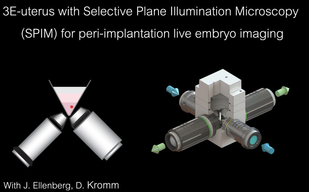
Biomechanics of embryo implantation
Implantation into the uterus redefines the physical context for the developing embryo. I am interested in elucidating the role of biomechanics in embryo-uterine coordination and development.
Using bioengineering, quantitative live imaging, and physical theory, I approached embryo implantation as a quantitative biomechanical phenomenon, allowing the understanding of the embryo-uterine interaction mechanisms via a theoretical physics framework of droplet wetting. In particular, I found that the integrin-mediated adhesion by the mTE provides the mechanism of TE tension release, driving morphogenesis of the pTE and ExE within the uterine geometric tissue context. The uterine tissue geometry spatially coordinates collective trophoblast migration to delineate space for egg cylinder growth.
Applying 3E-uterus revealed a new mechanism of coordination between extra-embryonic and embryonic tissue morphogenesis by the uterine microenvironment.
Publication
Bondarenko V, Nikolaev M, Kromm D, Belousov R, Wolny A, Blotenburg M, Zeller P, Rezakhani S, Hugger J, Uhlmann V, Hufnagel L, Kreshuk A, Ellenberg J, van Oudenaarden A, Erzberger A, Lutolf MP, Hiiragi T (2023).
Embryo-uterine interaction coordinates mouse embryogenesis during implantation.EMBO J, 42 (issue 17): e113280. (Cover article)
Collaborations
Dr. A. Erzberger, Dr. R. Belousov; EMBL Heidelberg
Prof. Dr. M. Lutolf, Dr. M. Nikolaev; EPFL/IHB Roche
Previous Research
I’ve made a transition to experimental research from initially computational training. Between 2013 and 2017, I performed my research in gene regulation, population, and comparative genomics.
Gene regulation during development
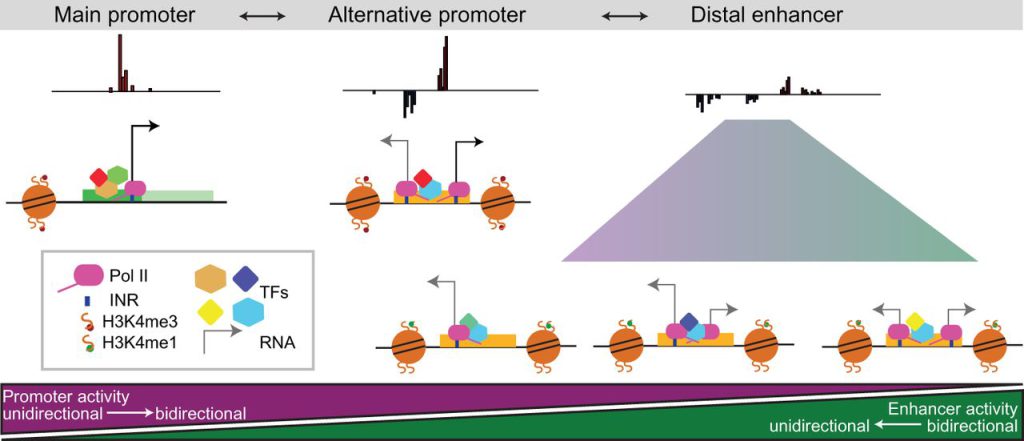
The initiation and regulation of transcription are attributed to promoters and enhancers, respectively, but both regulatory elements are transcriptionally active. In my M.Sc. study, I addressed the functional activity of enhancers and promoters in relation to their transcriptional properties.
Using bioinformatics and computational data analysis, I studied the genome-wide relationship between the regulatory activity of promoters and enhancers during Drosophila development and their transcriptional properties, such as the levels and the directionality of enhancer RNA (eRNA) transcription.
This study suggested the ‘continuum’ model of cis-regulation, where the balance between enhancer and promoter activity is generally reflected in the levels and directionality of eRNA transcription and is likely an inherent sequence property of the regulatory element.
Publication
Mikhaylichenko, O*, Bondarenko V*, Harnett D*, Schor IE, Males M, Viales RR, Furlong EE (2018). The degree of enhancer or promoter activity is reflected by the levels and directionality of eRNA transcription. Genes & Development. 32 (issue 1): 42-57. (Cover article)
* These authors contributed equally to this work
Online
R-loops in the regulatory genome
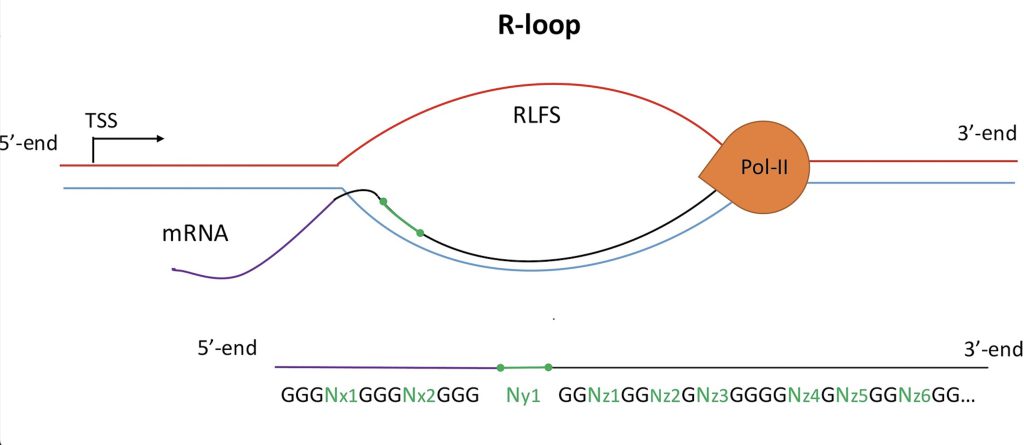
R-loops are three-stranded RNA:DNA hybrid structures that form during transcription.
I conducted a comprehensive comparative analysis of R-loop forming DNA regions with the experimental functional genomics data. Using statistics and bioinformatics tools, I characterized the genome-wide association of R-loop forming DNA sequences with gene promoters, U1 splice sites, gene ends, enhancers, and non-B DNA structures, such as G-quadruplexes. The co-localization of R-loop forming DNA sequences with promoters and transcriptionally active enhancers suggested new models for in cis and in trans regulation of transcription initiation by RNA:DNA hybrids.
This study provides a rationale and a computational prediction for the experimental study of the non-B DNA regulatory structures involved in the formation of the RNA:DNA interactome.
Publication
Kuznetsov VA, Bondarenko V, Wongsurawat T, Yenamandra SP, Jenjaroenpun P (2018). Toward predictive R-loop computational biology: genome-scale prediction of R-loops reveals their association with complex promoter structures, G-quadruplexes and transcriptionally active enhancers. Nucleic Acids Research. 46 (15): 7566-7585.
Population and comparative genomics
Bergman CM, Han S, Nelson MG, Bondarenko V, Kozeretska IA (2017). Genomic analysis of P elements in natural populations of Drosophila melanogaster. PeerJ. 15.5: e3824.
Online
Bondarenko V, Gelfand MS (2016). Evolution of the exon-intron structure in Ciliates. PLOS One. 11(9): e0161476.
Online
Impressum and Intellectual Property Clause
This website and its original content, features, and functionality are owned by Vladyslav Bondarenko and are protected by international copyright, trademark, patent, trade secret, and other intellectual property or proprietary rights laws.
One must not under any circumstances:
– Modify any materials and their copies from this website without formal approval from the owner (Vladyslav Bondarenko).
– Use any illustrations, images, photos, video or audio sequences or any graphics separately from the accompanying text, including original captions on the website. The source (Vladyslav Bondarenko & other involved and indicated persons) must always be acknowledged.
– Delete or alter any copyright, trademark, or other proprietary rights notices from copies of materials from the website.